


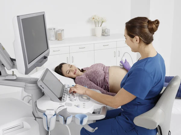
SonoCiné Automated Whole Breast Ultrasound (AWBUS) is a radiation-free imaging method that produces detailed images of breast tissue without compression. Knowing how the ultrasound works, how to prepare, and what to expect during the procedure can make patients more informed and comfortable. Here are answers to some frequently asked questions about the SonoCiné ultrasound experience:
How Does SonoCiné Work?
SonoCiné uses an automated arm to guide a handheld probe across the breast surface. The probe emits sound waves, and those waves bounce back to form detailed images. A technologist oversees the equipment throughout, so the scan follows a consistent approach across the tissue. The ultrasound system captures thousands of images as it moves, and it records them in a sequence a radiologist can review later. This automated approach reduces some of the variability associated with manual scanning, supporting thorough coverage. Dense breast tissue can obscure details on a mammogram, and ultrasound offers a way to see beyond that limitation.
Who Can Benefit?
Ultrasound screening suits several groups of women, and knowing where you fit can guide your decision. The technology works alongside other imaging methods, so it often serves as a helpful complement rather than a replacement. Here are common reasons women choose an automated ultrasound:
- Dense breast tissue: Mammograms may struggle to show clear results in dense breast tissue, and ultrasound provides a complementary view.
- Inconclusive mammogram results: A doctor may recommend further imaging when a mammogram raises questions.
- A desire for thorough screening: Some women want more information about their breast health.
- Sensitivity to compression: Ultrasound does not require the compression used in a mammogram.
These factors highlight why automated ultrasound can be a valuable addition to breast health screening.
What Happens During Screening?
The appointment follows a simple and predictable structure. You lie on a table, and the technologist applies a warm gel to the skin. This gel helps the sound waves travel, so the images come through clearly. The automated arm then moves the probe across each breast in sections. You stay still during the scan, though you can breathe normally throughout. The technologist monitors the equipment and adjusts the position as needed. To make sure your visit goes smoothly, keep the following preparation tips in mind:
- Wear a two-piece outfit: You’ll undress from the waist up, so separate pieces make it easier.
- Skip lotion or powder on the day: These products can interfere with the gel and sound waves.
- Bring prior imaging if available: Past results provide the radiologist with useful context.
- Arrive a few minutes early: Extra time lets you complete any paperwork without rushing.
These steps set you up for a straightforward appointment and reduce the risk of delays.
When Are Results Available?
A radiologist reviews the recorded images after your scan, and this review does not happen in the room with you. The specialist studies the full sequence of images, then prepares a report. Timing varies by facility; some centers deliver results within a few business days. Your provider then explains the findings, and you can ask questions during that conversation. The report may confirm a clear result or suggest the need for more imaging. If the radiologist recommends a follow-up, that step is a routine part of thorough screening. More imaging provides your care team with more detail and helps them reach a confident conclusion.
Schedule Your Sonociné Ultrasound Appointment
Sonociné ultrasound offers a comprehensive and precise supplementary screening method. It is designed to detect potential abnormalities that may be missed by traditional methods such as mammography. It enhances accuracy and supports a thorough evaluation of breast health by providing a painless and radiation-free process. Contact a reputable ultrasound clinic near you to schedule your sonociné ultrasound appointment today.
